

By Leigh Doyle
What started as a challenge between three graduate students in 1996 could today be a potential breakthrough for cancer treatment and organ transplants.
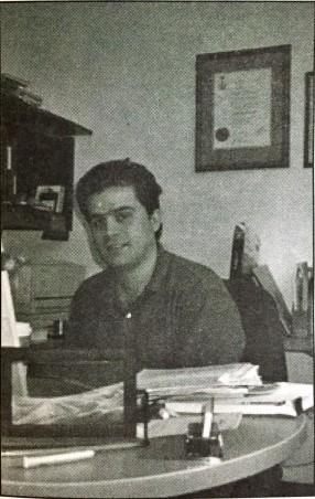
Ryerson assistant professor Michael Kolios has discovered that high frequency ultrasound can image apoptosis — the scientific name for the self-destruction of cells.
Over six years ago, Kolios and then fellow graduate student Gregory Czarnota attended a lecture on imaging thermal lesions. They asked the presenter, also a grad student, if this technique could be applied to image apoptosis. The answer was no. So Kolios and Czarnota set out to prove the presenter wrong.
During this time, Kolios attended the University of Toronto, where he completed his undergraduate degree in physics and PhD in medical biophysics — more specifically, heat transfer in tissue.
A nice offer brought him to Ryerson. Here, he is an associate professor with the math, physics and computer science department — teaching physics to first and second-year engineering and biology students.
High frequency ultrasound imaging is the focus of Kolios’ research. Much like an ultrasound done during pregnancy, this technique may be used to create an image of cells, revealing those that are becoming cancerous.
Cancer is physically represented by a malignant growth, but stems from an abnormal and uncontrolled division of body cells.
Currently, cancer patients must undergo treatment like chemotherapy for weeks before a doctor can check to see if it is working. To test the effectiveness of the treatment, a piece of the cancerous growth must be cut out (called a biopsy). Doctors can then see if treatment needs to be changed and the process starts again.
Kolios’ research team has successfully imaged apoptosis in artificial cell systems. The next step is to test this on humans who are currently receiving cancer treatments. By tracking apoptosis, doctors can see if a treatment is effective in destroying cancer cells. Kolios wants to image the cancerous cells before and after patients receive therapy.
“No one has ever seen this before,” Kolios said. If research and trials are successful, this ultrasound imaging would be the first non-invasive method to check cancer treatment.
“I’ve been extremely lucky. I’ve had access at the Princess Margaret hospital,” Kolios said. The ultrasound imaging device is located at Princess Margaret, and was purchased with a grant form the Canada Foundation for Innovation won by Kolios and his colleagues.
One of those colleagues, associate professor Bill Whelan, is working to step in after Kolios’ innovations open the door. Whelan works with Kolios, focusing his research on thermal therapy.
The idea of thermal therapy is to heat cancer cells with sources like lasers to temperatures higher than 55oC — which kills the cancerous cells. The challenge of the research, Whelan said, is to localise the heating to cancer cells alone without damaging healthy cells.
High frequency ultrasound imaging is useful to this research because it may be able to give a picture of the cells that have been damaged by heating.
Should both professors be successful, doctors could non-invasively test treatments and alter them early, without having to operate.
This would not only help customize patient treatment, but may spare some side effects on patients.
“It may directly impact be quality of cancer treatments,” Whelan said. He described Kolios as a dedicated researcher and his work as innovative. He said the research is very well received by both the international research community and Ryerson.
“Ryerson has been very supportive of the research,” Whelan said.


Leave a Reply